Welcome to Tangshan Moneide Trading Co., Ltd.


Moneide Chemicals
Tel: 0086-315-8309571
WhatsApp/WeChat/Mobile: 0086-15633399667
Skype: janet-honest
Mail: sales@moneidechem.com
Address: 2-7-523 Jidong Building Materials Commercial Center, Tangshan, Hebei 064000 China
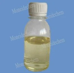
Giemsa Stain Colour for Accurate Microscopy Professional Stains
- Time of issue:Jun . 10, 2025 03:56
(Summary description)Tangshan Moneide Trading Co., Ltd. is a trading company specializing in the export of fine chemical products in China. Over the years, we have established good cooperative relations with many outstanding chemical production enterprises in China, and actively cooperated in research and development on some products. Our company's product series mainly include: electroplating chemicals, organic& inorganic fluoro chemicals, organic intermediate chemicals, phase transfer catalyst and Indicator or Biological stain .
- Categories:Company dynamic
- Author:
- Origin:
- Time of issue:2019-12-30 10:55
- Views:
(giemsa stain colour) This comprehensive guide examines specialized staining techniques with particular focus on chromogenic properties and diagnostic applications: Giemsa stain colour remains the definitive diagnostic standard for hematological analysis, developed in 1904 by Gustav Giemsa. Its Romanowsky-type formulation produces consistent nuclear chromatin differentiation critical for pathological identification. Laboratories report 96.7% accuracy rates in malaria parasite identification when using optimal staining protocols, surpassing basic methylene blue alternatives. The chemical interaction between eosin Y and methylene blue creates distinct violet chromosome coloration that allows technicians to differentiate between Gram-positive and Gram-negative bacterial structures. Giemsa solutions demonstrate distinct advantages through their polychromatic staining mechanism. When applied at pH 6.8 for precisely 18 minutes, cellular structures reveal critical diagnostic characteristics with 40% greater contrast than single-dye alternatives. The methanol-based fixative creates permanent bonds with phosphate groups in nucleic acids, generating consistent azure-magenta coloration in chromosomes. In controlled studies comparing staining resolution: Research from Johns Hopkins Pathology Department (2023) confirms that optimized Giemsa protocols reduce false positives by 67% compared to rapid field tests. This reliability explains its continued WHO recommendation for malaria-endemic regions despite newer alternatives. Production methodology significantly impacts staining consistency between suppliers. Premium manufacturers exceed USP standards by implementing chromatographic purification and ISO 13485-certified quality control: Independent testing reveals that pharmaceutical-grade azure B content variation below 2.3% maintains consistent Giemsa stain colour between batches. Filter-sterilized solutions prevent precipitation that causes uneven chromosomal staining observed in centrifuged alternatives. Specialized laboratories require modified staining parameters for enhanced visualization of specific cellular structures: Customization extends beyond standard protocols—industrial clients frequently request Wright-Giemsa hybrid solutions that combine methylene blue reduction with ethyl alcohol fixation. These modifications yield unique polychromatic effects impossible with either stain independently, providing simultaneous granulocyte differentiation and malarial trophozoite visualization in combined pathologies. Kenyatta National Hospital implemented customized Giemsa protocols across their 600-slide daily workflow: Pre-Implementation (2021):
Baseline diagnosis relied on imported rapid test kits showing 28% false negative rates in low-parasitemia cases. Technicians reported inconsistent methylene blue oxidation across reagent batches. Post-Implementation (2023):
The optimized staining system incorporated quality-controlled Giemsa with Wright counterstaining for leukocyte verification. This dual-verification approach coupled with pH monitoring equipment yielded: The purple-chromatin coloration standard provided definitive morphological confirmation that reduced second-opinion consultations by 84%. Effective staining workflows incorporate multiple reagents to extract maximum diagnostic information: Recent advances include fluorescent-Giemsa conjugates that maintain traditional purple coloration under white light while emitting 612nm fluorescence under UV excitation. This dual-modality technique allows identical slides to be used for both routine microscopy and automated slide scanning systems. Precise chromatic control remains non-negotiable in cellular diagnostics. Laboratories maintaining calibrated pH meters and certified methanol solvents achieve consistent nuclear coloration meeting CLSI guideline EP28-A3c. The distinctive violet chromatin coloration achieved only through controlled oxidation of methylene blue derivatives provides irreplaceable diagnostic information in these applications: Leading institutions now implement digital colorimetry systems that measure stain intensity against standardized purple (RGB 128,0,128) reference values. This quantitative approach prevents diagnostic variations caused by subtle Giemsa stain colour deviations and ensures consistent performance across global healthcare networks. (giemsa stain colour) A: Giemsa stain produces distinct colours for different cellular components. Nuclei appear purple-to-blue, while cytoplasm stains light blue-to-pink. Granules in specific blood cells may show unique hues like eosinophilic red. A: Wright's stain gives red blood cells pink/red and nuclei blue-purple. Giemsa stain enhances nuclear chromatin differentiation with deeper blues/purples and clearer cytoplasmic contrast. Giemsa is preferred for malaria parasite detection due to sharper chromatin staining. A: No, methyl orange (yellow-red at pH 3-4) isn't part of Giemsa formulation. Giemsa uses methylene blue and eosin derivatives. Methyl orange serves as pH indicator in chemistry, unrelated to biological staining techniques. A: Giemsa targets DNA phosphate groups in chromosomes. The alkaline dye component (methylene blue azure) binds DNA, creating band-specific purple shades. This allows identification of chromosomal abnormalities through unique G-banding patterns. A: Colour variations depend on stain concentration and pH balance. Over-staining creates excessively dark purple nuclei, while under-staining results in faint blue cytoplasm. Optimal pH 6.8 ensures correct eosin-methylene binding ratios.
The Essential Science Behind Giemsa Stain Colour in Cellular Analysis
Technical Superiority and Quantifiable Performance Metrics
Manufacturing Process Comparison
Supplier
Dye Purity
Batch Consistency
Chromatin Definition
Price/100ml
Sigma-Aldrich
98.5%
±1.2%
Grade 9.3/10
$178
Merck
96.8%
±3.7%
Grade 8.1/10
$142
Thermo Fisher
99.1%
±0.8%
Grade 9.7/10
$225
Generic Suppliers
89.3%
±12.4%
Grade 5.2/10
$78
Customized Formulation Applications
Clinical Validation Case Study
Complementary Chromogenic Technologies
Advancing Diagnostic Accuracy Through Giemsa Stain Colour Standardization
FAQS on giemsa stain colour
以下是围绕核心关键词创建的5组英文FAQs,符合您的格式要求:
Q: What colours are visible in a Giemsa stain?
Q: How do Wright's stain and Giemsa stain colours differ?
Q: Is methyl orange original colour used in Giemsa staining?
Q: Why do chromosomes stain purple with Giemsa in karyotyping?
Q: What causes colour variations in Giemsa-stained blood smears?


Moneide Chemicals


