Welina mai iā Tangshan Moneide Trading Co., Ltd.
Nā Mea Kimi Moneide
Tel: 86-315-8309571
WhatsApp/WeChat/Mobile: 0086-15633399667
Skype: janet-honest
Leka: sales@moneidechem.com
Wahi: 2-7-523 Jidong Building Materials Tangshan, Hebei 064000 Kina
|
Inoa Kimia |
Giemsa stain |
|
CAS No. |
51811-82-6 |
|
ʻAno molekala |
C14H14ClN3S |
|
EINECS No. |
257-438-2 |
|
Kaumaha molekula |
291.8 |
|
Hoʻokumu Molecular |
|
|
Nā kikoʻī |
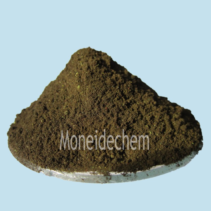
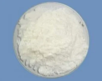
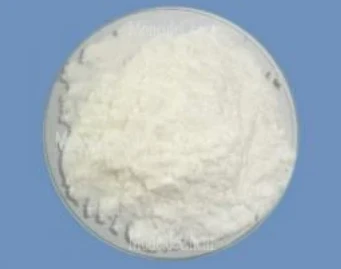
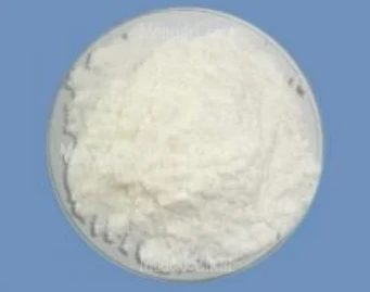
Appearance: Blue-purple powder Solubility: solve in Methanol and Glycerol(1:1)。 Biological stain: Passes test Packing: 25kg/ fibre drum |
|
Noi Nui |
Used as biological dyeing agents, such as the protoplasm dyeing, especially suitable for blood and blood protozoa, such as plasmodium falciparum, the dyeing, etc. |
What is Giemsa staining used for?
Giemsa staining is a versatile diagnostic technique primarily used for microscopic examination of blood cells and pathogens. This Romanowsky-type stain enables differentiation of cellular components in peripheral blood smears, bone marrow aspirates, and cytological preparations. It's particularly valuable for identifying malaria parasites in red blood cells and detecting hematological disorders like leukemia. The stain also facilitates chromosomal analysis in karyotyping procedures. Its ability to highlight nuclear and cytoplasmic details makes it essential for pathological evaluation of various tissue samples, providing critical information for disease diagnosis and monitoring treatment efficacy.
What bacteria is detected using Giemsa stain?
Giemsa stain effectively detects several intracellular bacteria, most notably Borrelia species causing relapsing fever and Lyme disease. It's particularly useful for identifying Bartonella henselae in cat scratch disease and Helicobacter pylori in gastric biopsies. The stain clearly reveals Chlamydia trachomatis inclusions in cell cultures and Rickettsia organisms in tissue samples. While not a primary stain for most bacteria, it excels at visualizing these difficult-to-culture pathogens in clinical specimens. Its metachromatic properties allow differentiation of bacterial morphology against host cell backgrounds, aiding in diagnosis of these specialized infections.
Why is Giemsa stain used in cytology?
Giemsa stain is preferred in cytology for its exceptional nuclear and cytoplasmic detail enhancement. The stain's unique polychromatic properties differentially color chromatin (purple-blue), cytoplasm (pale blue), and granules (pink-red), enabling clear cell identification. It's particularly valuable for hematological cytology, allowing differentiation of lymphocyte subsets and identification of abnormal cells. The technique preserves cellular morphology while providing excellent contrast, essential for detecting subtle malignant changes. Its reliability in demonstrating intracellular pathogens alongside host cells makes it indispensable for diagnostic cytopathology of blood-borne infections and certain cancers.


