મોનેઇડ કેમિકલ્સ
Tel: 86-315-8309571
WhatsApp/WeChat/Mobile: 0086-15633399667
સ્કાયપે: જેનેટ-પ્રામાણિક
મેઇલ: sales@moneidechem.com
સરનામું: 2-7-523 જીડોંગ બિલ્ડિંગ મટિરિયલ્સ તાંગશાન, હેબેઈ 064000 ચીન
|
Chemical Name |
Wright's stain |
|
CAS નં. |
68988-92-1 |
|
પરમાણુ સૂત્ર |
C36H27N3O5Br4S |
|
EINECS નં. |
273-541-5 |
|
પરમાણુ વજન |
—— |
|
પરમાણુ માળખું |
|
|
વિગતો |
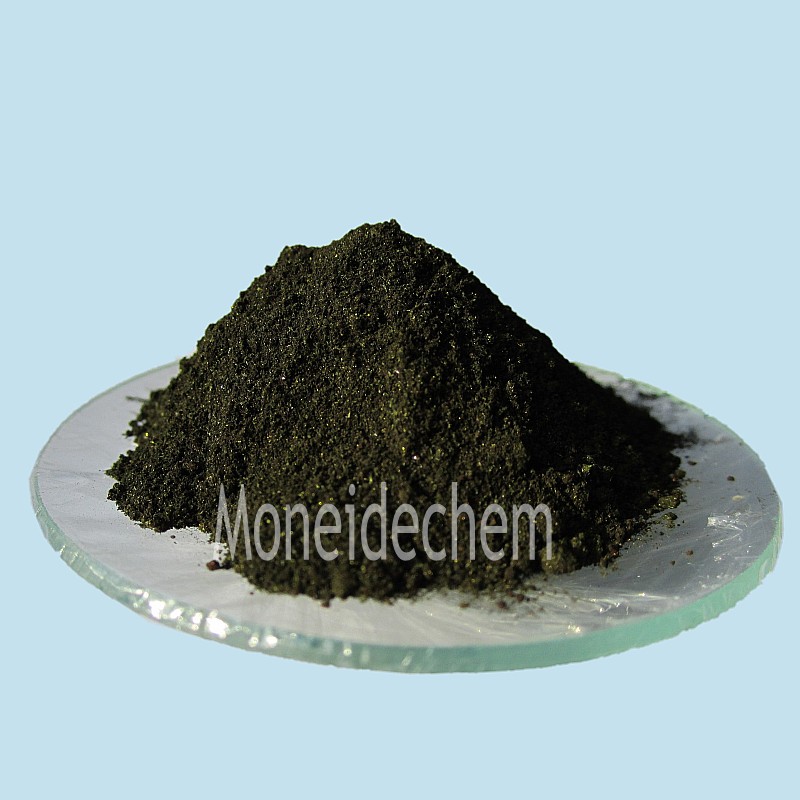
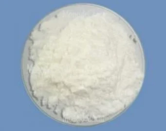
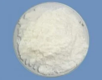
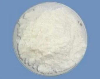
Appearance: Bright green powder Solubility: insolve in water,solve in methanol and ethanol Biological stain: Passes test Packing: 25kg/ fibre drum |
|
મુખ્ય એપ્લિકેશન |
Used as biological dyeing agents. |
1. What is Wright's stain?
Wright's stain is a polychromatic dye used primarily in hematology and histology to differentiate blood cell types under a microscope. It consists of a mixture of eosin (an acidic dye) and methylene blue (a basic dye), which react with cellular components to produce varying colors. The stain highlights nuclei in purple-blue, cytoplasm in pink, and granules in specific hues, aiding in identifying white blood cells, red blood cells, and platelets. It is commonly employed for peripheral blood smears and bone marrow samples, providing critical diagnostic information for conditions like anemia, infections, and leukemia.
2. What is the difference between Wright's stain and Giemsa stain?
Wright's stain and Giemsa stain are both Romanowsky stains but differ in composition and application. Wright's stain uses eosin and methylene blue dissolved in methanol, offering rapid staining ideal for clinical blood smears. Giemsa stain incorporates azure B and eosin Y, requiring a buffer for optimal pH, and is preferred for detecting parasites like malaria or detailed cytogenetic studies. While Wright's stain provides quicker results, Giemsa offers superior nuclear and cytoplasmic contrast, especially for protozoan and bacterial identification in thicker specimens.
3. What is the Wright's stain used for bacteria?
Wright's stain is occasionally used for bacterial visualization but is less common than Gram or Giemsa staining. It can highlight certain bacteria in blood smears or tissue sections by imparting a purple-blue color to their nucleic acids. However, it lacks specificity for bacterial classification (e.g., Gram-positive vs. Gram-negative) and is primarily reserved for intracellular pathogens like Rickettsia or Anaplasma within blood cells. Its utility for bacteria is limited compared to dedicated bacterial stains, as it prioritizes eukaryotic cell differentiation over prokaryotic detail.


